NERVOUS SYSTEM
Components : brain , spinal cord, nerves and special sense organs (e.g., eyes and ears).
· Generates nerve impulses (action potentials) to regulate body activities.
· Detects changes in the body’s internal and external environment.
· Interprets the changes and responds by causing muscular contractions or glandular secretions.
BRAIN


FUNCTIONS:


BRAIN

CEREBRUM

1.INTRODUCTION :- it is largest part of the brain .
2.SITUATION : it is situated in the cranium occupies the anterior and middle cranial fossae.
3.CEREBRUM is divided by a deep cleft,the longitudinal cerebral fissure, into right and left cerebral hemispheres, each containing one of the lateral ventricles.


- Deep with in the brain the hemisphers are connected by a mass of white matter(nerve fibers) called the “corpus callosum”.
- The falx cerebri is formed by the dura mater.
- It separates the two hemispheres and penetrates to the depth of the “corpus collasum”
- The superficial part of the cerebrum is composed of nerve cell bodies (or) Grey Matter forming the cerebral cortex.
- The deeper layers consist of nerve fibers or white matter.
- The cerebral cortex shows many infoldings or furrows of varying depth .
- The exposed areas of the folds are gyri or convolutions greatly increase the surface area of the cerebrum
3) PARTS
Divided into lobes with the names of the bones of the cranium.

1.FRONTAL
2.PARIETAL
3.TEMPORAL
4.OCCIPITAL
The boundaries of the lobes are marked by deep sulci.

INFERIOR OF THE CEREBRUM:-
The surface of the cerebral cortex is composed of grey matter (nerve cell bodies )
Within the cerebrum the lobes are connected by mass of nerve fibers, or tracts which make up the white matter of the brain .
The efferent and afferent fibers linking the different parts of the brain and spinal cord as follows.
1. ASSOCIATION(ACRUATE) TRACTS
2. COMMISSURAL TRACTS
3. PROJECTION TRACTS
ASSOCIATION TRACTS:-
It connect different parts of a cerebral hemispheres by extending from one gyrus to another.
COMMISSURAL TRACTS:-
It connects corresponding areas of the two hemisphere by extending from gyrus to another.
It mainly maintains the “corpus callosum”.
PROJECTION TRACTS:-
It connects the cerebral cortex with grey matter of lower parts of the brain and with the spinal cord, example the internal capsule.
THE INTERNAL CAPSULE :-
It is an important projection tract that lies deep within the brain between the basal ganglia (nuclei) and the thalamus.
FUNCTIONS OF THE CEREBRAL CORTEX

Functional areas of the cerebral cortex.
Motor areas of the cerebral cortex.
Sensory areas of the cerebral cortex.
Association areas.
Other areas of the cerebrum .
THALAMUS

v The thalamus consist of large masses of grey matter.
v It contains many nuclei.
v It is predominantly a sensory relay station with incoming fibres from the spinal cord and brain stem and onward fibres to the cerebral cortex.
v Thethalamus is the crude identification of stimuli such as pain.
v Variations of temperature (or) touch is due to thalamic integration.
HYPOTHALAMUS:
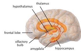
v It lies below the thalamus.
v Various afferent and efferent connections of hypothalamus are responsible for its functions.
v It is having connections with limbic system.
v Nuclei of tegmentum ,pons,hind brain and with pituitary gland.
FUNCTIONS:
v TEMPERATURE REGULATION: normal body temperature is maintained by striking a balance between heat production and heat loss.
1. Temperature regulating centre in the hypothalamus is sensitive to changes in temperature of blood and also receives input from nerve fibres innervating temperature receptors in the skin.
2. The hypothalamus regulates the secretion of the anterior and posterior pituitary hormones.
3. Regulation of body water and electrolyte concentration .
4. Maintenance of normal sexual desire, behaviour and reproduction.
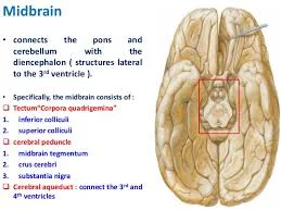
MIDBRAIN –(MESENCEPHALON):

1. It is the shortest segment of the brain stem, joins the forebrain above to the pons and cerebellum below.
2. Mid brain divided into right and left cerebral peduncles .

3. Two ventral portions of these are distinctly separate from each other called CRURA CEREBRI.
A LAYER OF GREY MATTER

1. A layer of grey matter –substantia nigra seprates them from the dorsal part of the midbrain, which is continues across the midline is known as TEGMENTUM.

2. The matter is transversed by the cerebral aqueduct which connects the 3rd and 4th ventricles.
3. The part of the tegmentum dorsal to the aqueduct is called tectum and it presents four rounded elevations known as the COLLICULI.
4. The upper pair super colliculi are visual reflex centres and the lower pair super colliculi are visual reflex centres and the lower pair inferior colliculi are auditory reflex centres.
5. Mid brain is an important centre for various righting and postural reflexes.
6. These reflexes are being directed through visual and auditory impulses.


OTIC GANGLION

INTRODUCTION :
It is a peripheral parasympathetic ganglion which relays secretomoter fibres to the parotid gland.
Topographically , it is intimately related to the mandibular nerve , but functionally it is a part of the glossopharyngeal nerve.
SIZE AND SITUATION
a) It is 2-3 mm in size , and is situated in the infratemporal fossa ,just below the foramen ovale.
b) It lies medial to the mandibular nerve , and lateral to the tensor veli palatine.
c) It surrounds the origin of the nerve to the medial pterygoid.
CONNECTIONS AND BRANCHES

a) The motor or parasympathetic root is formed by the lesser petrosal nerve.
b) The preganglionic fibres are derived from the inferior salivary nucleus-the ninth nerve, its tympanic branch, the tympanic plexus –the lesser petrosal nerve to reach the ganglion.
c) The postganglionic or secretomotor fibres pass through the auriculotemporal nerve.they are vasomotor in function .
d) The sensory root comes from the aurioculotemporal nerve and is sensory to the parotid gland.
Other fibres passing through the ganglion are as follows.
a) The nerve to the medial pterygoid gives a motor root to the ganglion which passes through it without relay and supplies medially placed tensorveli palatini and laterally placed tensor tympani muscles.
b) The chorda tympani nerve is connected to the otic ganglion and also to the nerve of the pterygoid canal. These connectins provide an alternative pathway of taste from the anterior two-thirds of the tongue. These fibres do not pass through the middle ear.
. 8 cervical
PERIPHERAL NERVOUS SYSTEM
This part of the nervous system consists of:
· . 31 pairs of spinal nerves
· . 12 pairs of cranial nerves
· . the autonomic nervous system.
Most of the nerves of the peripheral nervous system are composed of sensory nerve fibres conveying afferent impulses from sensory organs to the brain, and motor nerve fibres conveying efferent impulses from the brain to the effector organs, e.g. skeletal muscles, smooth muscle and glands.
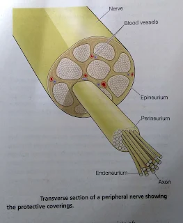
Each nerve consists of numerous nerve fibres collected into bundles.
Each bundle has several coverings of protective connective tissue.

· . Endoneurium is a delicate tissue, surrounding each individual fibre, which is continuous with the septa that pass inwards from the perineurium.
· . Perineurium is a smooth connective tissue, surrounding each bundle of fibres.
· . Epineurium is the fibrous tissue which surrounds and encloses a number of bundles of nerve fibres. Most large nerves are covered by epineurium.
HIT LIKE AND SHARE
Spinal nerves


There are 31 pairs of spinal nerves that leave the vertebral canal by passing through the intervertebral foramina formed by adjacent vertebrae.
They are named and grouped according to the vertebrae with which they are associated:
They are named and grouped according to the vertebrae with which they are associated:
. 8 cervical
. 12 thoracic
. 5 lumbar
. 5 sacral
. 1 coccygeal.
Although there are only seven cervical vertebrae, there are eight nerves because the first pair leaves the vertebral canal between the occipital bone and the atlas and the eighth pair leave below the last cervical vertebra.
Thereafter the nerves are given the name and number of the vertebra immediately above.
Thereafter the nerves are given the name and number of the vertebra immediately above.
The lumbar, sacral and coccygeal nerves leave the spinal cord near its termination at the level of the first lumbar vertebra, and extend downwards inside the vertebral canal in the subarachnoid space, forming a sheaf of nerves which resembles a horse’s tail, the cauda equine.
These nerves leave the vertebral canal at the appropriate lumbar, sacral or coccygeal level, depending on their destination.
These nerves leave the vertebral canal at the appropriate lumbar, sacral or coccygeal level, depending on their destination.
Nerve roots
The spinal nerves arise from both sides of the spinal cord and emerge through the intervertebral foramina.
Each nerve is formed by the union of a motor (anterior) and a sensory (posterior) nerve root and is, therefore, a mixed nerve.
Thoracic and upper lumb ar (L1 and L2) spinal nerves have a contribution from the sympathetic part of the autonomic nervous system in the form of a preganglionic fibre.
Bones and joints are supplied by adjacent nerves.
The anterior nerve root consists of motor nerve fibres, which are the axons of the lower motor neurons from the anterior column of grey matter in the spinal cord and, in the thoracic and lumbar regions, sympathetic nerve fibres, which are the axons of cells in the lateral columns of grey matter.
The posterior nerve root consists of sensory nerve fibres. Just outside the spinal cord there is a spinal ganglion (posterior root ganglion), consisting of a little cluster of cell bodies.
Sensory nerve fibres pass through these ganglia before entering the spinal cord.
The area of skin whose sensory receptors contribute to each nerve is called a dermatome.
Sensory nerve fibres pass through these ganglia before entering the spinal cord.
The area of skin whose sensory receptors contribute to each nerve is called a dermatome.
For a very short distance after leaving the spinal cord the nerve roots have a covering of dura and arachnoid maters.
These terminate before the two roots join to form the mixed spinal nerve. The nerve roots have no covering of pia matter.
These terminate before the two roots join to form the mixed spinal nerve. The nerve roots have no covering of pia matter.
Branches
Immediately after emerging from the intervertebral foraminen, spinal nerves divide into brances, or rami: a ramus communicans, a posterior ramus and an anterior ramus.
The rami communicante are part of preganglionic sympathetic neurons of the autonomic nervous system.
The posterior rami pass backwards and divide into medial and lateral branches to supply skin and muscles of relatively small area of the posterior aspect of the head, neck and trunk.
The anterior rami supply the anterior and lateral aspects of the neck, trunk, and upper and lower limbs.


The femoral nerve is one of the larger branches. It passes behind the inguinal ligament to enter the thigh in close association with the femoral artery.
CRANIAL NERVES

There are 12 pairs of cranial nerves originating from nuclei in the inferior surface of the brain, some sensory, some motor and some mixed. Their names and numbers are:
- I. Olfactory: sensory
- II. Optic: Sensory
- III. Oculomotor : motor
- IV. Trochlear: motor
- V: Trigeminal: mixed
- VI: Abducent: motor
- VII. Facial: mixed
- VIII. Vestibulocochlear (auditory): sensory
- IX. Glossophranyngeal: mixed
- X. Vagus: mixed
- XI. Accessory: motor
- XII. Hypoglossal: motor.
I. OLFACTORY NERVES (SENSORY)

- These are the nerves of the sense of smell.
- Their sensory receptors and fibers originate in the upper part of the mucous membrane of the nasal cavity, pass upwards through the cribriform plate of the ethmoid boned and then go to the olfactory bulb.
- The nerves then proceed backwards as the olfactory tract, to the area for the perception of smell in the temporal lobe of the cerebrum.
II. OPTIC NERVES (SENSORY)

- These are the nerves of the sense of sight.
- The fibres originate in the retinae of the eyes and they combine to form the optic nerves.
- They are directed backwards and medially through the posterior part of the orbital cavity. They then pass through the optic foramina of the sphenoid bone into the cranial cavity and join at the optic chiasma.
- The nerves proceed backwards as the optic tracts to the lateral geninculate bodies of the thalamus.
- Impulses pass from these to the centre for sight in the occipital lobes of the cerebrum and to the cerebellum.
- In the occipital lobe sight is perceived, and in the cerebellum the impulses from the eyes contribute to the maintenance of balance, posture and orientation of the head in space.
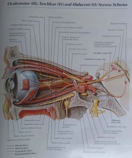
III. OCULOMOTOR NERVES (MOTOR)

- These nerves arise from nuclei near the cerebral aqueduct. They supply:
- . Four of the six extrinsic muscles, which move the eyeball, i.e. the superior, medial and inferior recti and the inferior oblique muscle.
- . the intrinsic (intraocular) muscles:
- - ciliary muscles, which alter the shape of the lens, changing its refractive power
- - circular muscles of the iris, which constrict the pupil
- . the levator palpebrae muscles, which rise the upper eyelids.
IV. TROCHLEAR NERVES (MOTOR)

- These nerves rise from nuclei near the cerebral aqueduct.
- They supply the superior oblique muscles of the eyes.
V. TRIGEMINAL NERVES (MIXED)

- These nerves contain motor and sensory fibers and are among the largest of the cranial nerves. They are the chief sensory nerves for the face and head (including the oral and nasal cavities and teeth), receiving impulses of pain, temperature and touch.
- The motor fibres stimulate the muscles of mastication.
- As the name suggests, there are three main branches of the trigeminal nerves.
- The dermatomes innervated by the sensory fibres on the right side.
- The ophthalmic nerves are sensory only and supply the lacrimal glands, conjunctiva of the eyes, forehead, eyelids, anterior aspect of the scalp and mucous membrane of the nose.
- The maxillary nerves are sensory only and supply the cheeks, upper gums, upper teeth and lower eyelids.
- The mandibular nerves contain both sensory and motor fibres.
- These are the largest of the three divisions and they supply the teeth and gums of the lower jaw, pinnae of the ears, lower lip and tongue.
- The motor fibres supply the muscles of mastication.
VI. ABDUCENT NERVES (MOTOR)
These nerves arise from nuclei lying under the floor of the fourth ventricle.
They supply the lateral rectus muscles of the eyeballs.
VII. FACIAL NERVES (MIXED)

These nerves are composed of both motor and sensory nerve fibres, arising from nuclei in the lower part of the pons.
The motor fibres supply the muscles o facial expression.
The sensory fibres convey impulses from the taste buds in the anterior two-thirds of the tongue to the taste perception area in the cerebral cortex.
VIII. VESTIBULOCOCHLEAR (AUDITORY) NERVES (SENSORY)

These nerves are composed of two distinct sets of fibres, vestibular nerves and cochlear nerves.
The vestibular nerves arise from the semicircular canals of the inner ear and convey impulses to the cerebellum.
They are associated with the maintenance of posture and balance.
The cochlear nerves originate in the spiral organ (of Corti) in the inner ear and convey impulses to the hearing areas in the cerebral cortex where sound is perceived.
IX. GLOSSOPHARYNGEAL NERVES (MIXED)

- The motor fibres arise from nuclei in the medulla oblongata and stimulate the muscles of the tongue and pharynx and the secretory cells of the parotid (salivary) glands.
- The sensory fibres convey impulses to the cerebral cortex from the posterior third of the tongue, the tonsils and pharynx and from taste buds in the tongue and pharynx.
- These nerves are essential for the swallowing and gag reflexes.
X. VAGUS NERVES (MIXED)

- These nerves have a more extensive distribution than any other cranial nerves.
- They pass down through the neck into the thorax and the abdomen.
- These nerves from an important part of the parasympathetic nervous system.
- The motor fibres arise from nuclei in the medulla and supply the smooth muscle and secretory glands of the pharynx, larynx, trachea, heart, oesophagus, stomach, intestines, exocrine pancreas, gall bladder, bile ducts, spleen, kidneys, ureter and blood vessels in the thoracic and abdominal cavities.
- The sensory fibres convey impulses from the membranes lining the same structures to the brain.
XI. ACCESSORY NERVES (MOTOR)

- These nerves arise from nuclei in the medulla oblongata and in the spinal cord.
- The fibres supply the sternocleidomastoid and trapezius muscles.
- Branches join the vagus nerves and supply the pharyngeal and laryngeal muscles.
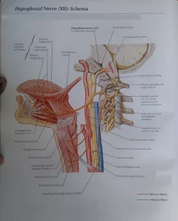
XII. HYPOGLOSSAL NERVES (MOTOR)

- These nerves arise from nuclei in the medulla oblongata.
- They supply the muscles of the tongue and muscles surrounding the hyoid bone and contribute to swallowing and speech.
Nerve roots
The spinal nerves arise from both sides of the spinal cord and emerge through the intervertebral foramina. Each nerve is formed by the union of a motor (anterior) and a sensory (posterior) nerve root and is, therefore, a mixed nerve. Thoracic and upper lumb ar (L1 and L2) spinal nerves have a contribution from the sympathetic part of the autonomic nervous system in the form of a preganglionic fibre.
the bones and muscles mentioned in the following section. Bones and joints are supplied by adjacent nerves.
the bones and muscles mentioned in the following section. Bones and joints are supplied by adjacent nerves.
The anterior nerve root consists of motor nerve fibres, which are the axons of the lower motor neurons from the anterior column of grey matter in the spinal cord and, in the thoracic and lumbar regions, sympathetic nerve fibres, which are the axons of cells in the lateral columns of grey matter.
The posterior nerve root consists of sensory nerve fibres. Just outside the spinal cord there is a spinal ganglion (posterior root ganglion), consisting of a little cluster of cell bodies. Sensory nerve fibres pass through these ganglia before entering the spinal cord. The area of skin whose sensory receptors contribute to each nerve is called a dermatome.
For a very shortdistance after leaving the spinal cord the nerve roots have a covering of dura and arachnoid maters. These terminate before the two roots join to form the mixed spinal nerve. The nerve roots have no covering of pia matter.
Branches
Immediately after emerging from the intervertebral foraminen, spinal nerves divide into brances, or rami: a ramus communicans, a posterior ramus and an anterior ramus.
The rami communicante are part of preganglionic sympathetic neurons of the autonomic nervous system.
The posterior rami pass backwards and divide into medial and lateral branches to supply skin and muscles of relatively small area of the posterior aspect of the head, neck and trunk.
The anterior rami supply the anterior and lateral aspects of the neck, trunk, and upper and lower limbs.
Plexuses
In the cervical, lumbar and sacral regions the anterior rami unite near their origins to form large masses of nerves, or plexuses, where nerve fibres are regrouped and rearranged before proceeding to supply skin, bones, muscles and joints of a particular area. This means that these structures have a nerve supply from more than one spinal nerve and therefore damage to one spinal nerve does not cause loss of function of a region.
In the thoracic region the anterior rami do not form plexuses.
There are five large plexuses of mixed nerves formed on each side of the vertebral column. They are the:
1. cervical plexuses
2. brachial plexuses
3. lumbar plexuses
4. sacral plexuses
5. coccygeal plexuses.
Cervical plexus
This is formed by the anterior rami of the first four cervical nerves. It lies opposite the 1st, 2nd, 3rd and 4th cervical vertebrae under the protection of the sternocleidomastoid muscle.
The superficial branches supply the structures at the back and side of the head and the skin of the front of the neck to the level of the sternum.
The deep branches supply muscles of the neck, e.g. the sternocleidomastoid and the trapezius.
The phrenic nerve originates from cervical roots 3, 4 and 5 and passes downwards through the thoracic cavity in front of the root of the lung to supply the diaphragm with impulses that stimulate contraction, initiating inspiration.
Brachial plexus
The anterior rami of the lower four cervical nerves and a large part of the first thoracic nerve from the brachial plexus shows its formation and the nerves that emerge from it. The plexus is situated in the neck and shoulder above and the behind the subclavian vessels and in the axilla.
The branches of the brachial plexus supply the skin and muscles of the upper limbs and some of the chest muscles. Five large nerves and a number of smaller ones emerge from this plexus, each with a contribution from more than one nerve root, containing sensory, motor and autonomic fibres.
. axillary (circumflex) nerve: C5, 6
. radial nerve: C5, 6, 7, 8, T1
. musculocutaneous nerve: C5, 6, 7
. median nerve: C5, 6, 7, 8, T1
. ulnar nerve: C7, 8, T1
. meidal cutaneous nerve: C8, T1.
The axillary (circumflex) nerve winds round the humerus at the level of the surgical neck. It then breaks up into minute branches to supply the deltoid muscle, shoulder joint and overlying skin.
The radial nerve is the largest branch of the brachial plexus. It supplies the triceps muscle behind the humerus, crosses in front of the elbow joint then winds round to the back of the forearm to supply extensors of the wrist and finger joints. It continues into the back of the hand to supply the skin of the thumb, the first two fingers and the lateral half of the third finger.
The musculocutaneous nerve passes downwards to the lateral aspect of the forearm. It supplies the muscles of the upper arm and the skin of the forearm.
The median nerve passes down the midline of the arm in close association with the brachial artery. It passes in front of the elbow joint then down to supply the muscles of the front of the forearm. It continues into the hand where it supplies small muscles and the skin of the front of the thumb, the first two fingers and the lateral half of the third finger. It gives off no branches above the elbow.
The ulnar nerve descends through the upper arm lying medial to the brachial artery. It passes behind the medial epicondyle of the humerus to supply the muscles on the ulnar aspect of the forearm. It continues downwards to supply the muscles in the palm of the hand and the skin of the whole of the little finger and the medial half of the third finger. It gives off no branches above the elbow.
The main nerves of the arm are presented. The distribution and origins of the cutaneous sensory nerves of the arm, i.e. the dermatomes, are shown.
Lumbar plexus
The lumbar plexus is formed by the anterior rami of the first three and part of the fourth lumbar nerves. The plexus is situated in front of the transverse processes of the lumbar vertebrae and behind the psoas muscle. The main branches, and their nerve roots are:
. iliohypogastric nerve: L1
. ilioinguinal nerve: L1
. genitofemoral: L1, 2
. lateral cutaneous nerve of thigh: L2, 3
. femoral nerve: L2, 3, 4
. obturator nerve: L2, 3, 4
. lumbosacral trunk: L4, (5).
The iliohypogastric, ilioinguinal and genitofemoral nerves supply muscles and the skin in the area of the lower abdomen, upper and medial aspects of the thigh and the inguinal region.
The lateral cutaneous nerve of the thigh supplies the skin of the lateral aspect of the thigh including part of the anterior and posterior surfaces.


0 Comments