LYMPH


LYMPH NODES

v Lymph is a clear watery fluid.
v Lymph is similar in composition to plasma,with the important exception of plasma proteins,and identical in composition to interstitial fluid.
v Lymph transports the plasma proteins that seep out of the capillary beds back to the blood stream.
v Lymph also carries away larger particles, e.g.bacteria and cell debris from damaged tissues,which can then be filtered out and destroyed by the lymph nodes.
v Lymph contains lymphocytes, which circulate in the lymphatic system allowing them to patrol the different regions of the body.
v In the lacteals of the small intestine,fats absorbed into the lymphatics give the lymph (chyle).
Chyle is a milky appearance.
LYMPH VESSELS.
LYMPH CAPILLARIES :

Ø These originate as blind-end tubes in the interstitial spaces.
Ø They have the same structures as blood capillaries,i.e. a single layer of endothelial cells,but their walls are more permeable to all interstitial fluid constituents ,including proteins and cell debris.
Ø The tiny capillaries join up to form larger lymph vessels.
Ø Nearly all tissues have a network have a network of lymphatic vessels,the exceptions being the central nervous system ,the bones and the most superficial layers of the skin.
LARGE LYMPH VESSELS .

1. The walls of lymph vessels are about the same thickness as those of small veins and have the same layers of tissue, i.e. a fibrous covering , a middle layer of smooth muscle and elastic tissue and an inner lining of epithelium .
2. Lymph vessels have numerous cup-shaped valves to ensure that lymph flows in one way only, i.e towards the thorax.
3. There is no pump,like the heart ,involved in the walls of the large lymph vessels has an intrinsic ability to contract rhythmically (the lymphatic pump).
4. Any structure that periodically compress es the lymphatic vessels can assist in the movement of lymph along the vessels,commonly including the contraction of adjacent muscles and the pulsation of large arteries.
5. Lymph vessels become larger as they join together,eventually forming two large ducts,the thoracic duct and right lymphatic duct, which empty into the subclavian veins.


THORACIC DUCT

v Thoracic duct begins at the cistern chili.
v Cisterna lymph channel situated in front of the bodies of the first two lumbar vertebrae.
v The duct is about 40 cm long and opens into the left subclavian vein in the root of the neck.
v It drains lymph from both legs ,the pelvic and abdominal cavities,the left half of the thorax,head and neck and the right arm.
RIGHT LYMPHATIC DUCT

· This is a dilated lymph vessel about 1 cm long.
· It lies in the root of the neck and opens into the right subclavian vein.
It drains lymph from the right half of the thorax,head and neck and the right armLYMPH NODES

1. INTRODUCTION:

Lymph nodes are oval or bean-shaped organs.
2. SITUTATION :
Lymph nodes are present in groups,along the length of lymph vessels.
3. SHAPE:
Oval or bean shape, size varies some are small as a pin head and the largest are about the size of an almond.
4. STRUCTURE :

v Lymph nodes have an outer capsule of fibrous tissue that dips down into the node substance forming partitions, or trabeculae.
v The main substance of the node consists of reticular and lymphatic tissue containing many lymphocytes and macrophages.
v As many as four or five afferent lymph vessels may enter a lymph node while only one efferent vessel carries lymph away from the node.
v Each node has a concave surface called the hilum, where an artery enters and a vein and the efferent lymph vessel leave.
v The large numbers of lymph nodes situated in strategic positions throughout the body are arranged in deep and superficial cervical needs.
v Lymph from the head and neck passes through deep and superficial cervical nodes.

v Lymph from the upper limbs passes through nodes situated in the elbow region,then through the deep and superficial axillary nodes.
v Lymph from organs and tissues in the thoracic cavity drains through groups of nodes situated close to the mediastenium,large airways,oesophagus and chest wall.

v Most of the lymph from the breast passes through the axillary nodes.

v Lymph from the pelvic and abdominal cavities passes through many lymph nodes before entering the cistern chyli.
5. BLOOD SUPPLY:

The abdominal and pelvic nodes are situated mainly in association with the blood vessels supplying the organs and close to the main arteries, i.e. the aorta and the external and internal iliac arteries.
6. FUNCTIONS:
Ø FILTERING AND PHAGOCYTOSIS .
§ Lymph is filtered by the reticular and lymphoid tissue as it passes through lymph nodes.
§ Particulate matter may include microbes,dead and alive phagocytes containing ingested microbes,cells from malignant tumours,worn-out and damaged tissue cells and inhaled particles.
§ Organic material is destroyed in lymph nodes by macrophages and antibodies.
§ In some cases where phagocytosis of microbes is incomplete they may stimulate inflammation and enlargement of node.(lymphoadenopathy).
Ø PROLIFERATION OF LYMPHOCYTES:
§ Activated T-and B-lymphocytes multiply in lymph nodes.
§ Antibodies produced by sensitised B-lymphocytes enter lymph and blood draining the node.

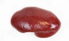

SPLEEN

1. Introduction : it is the largest lymph organ and formed by reticular and lymphoid
tissue.
2. Situation : it lies in the left hypochondriac region of the abdominal cavity
between the fundus of the stomach and the diaphragm.
3. Measurments : 1. Colour – purplish
2.Length – 12 cm long
3.Width – 7 cm diameter
4. thickness – 2.5 cm
5. weight - 200gm.
6. shape – slightly oval
4.
ORGANS ASSOCIATED WITH THE SPLEEN
Superiorly and posteriorely –
diaphragm
Inferiorly - left colic flexure of
the large intestine .
Anteriorly -
fundus of the stomach
Medially -
pancreas and the left kidney
Laterally - separated from the 9th
,10th and 11th ribs and the intercostal muscles by the diaphragm .
1.
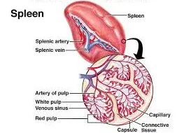
STRUCTURE
: the spleen is slightly oval in shape with the
hilum on the lower medial border .
1. The anterior surface is covered with peritoneum
.
2. It is enclosed in fibroelastic capsule that dips
into the organ, forming trabeculae.
2. The cellular material consisting of lymphocytes
and macrophages, is called splenic pulp , and it lies
between the trabeculae.
3. Red pulp is the part suffused with blood and
white pulp consists of areas of lymphatic tissue where there are sleeves of
lymphocytes and macrophages around blood vessels.
2.
BLOOD SUPPLY :
Arterial supply : splenic artery ,
a branch of the coeliac artery.
Venous drainage : splenic vein , a branch of
the portal vein.
Blood passing through the spleen flows in
sinuses which have distinct pores between the endothelial cells , allowing it
to come into close association with splenic pulp.
3.
NERVE SUPPLY - symphathetic nerve supply
Para
symphathetic nerve supply
8. FUNCTIONS : a. phagocytosis
b.
storage of blood
c. it
improves immune response
d. it
helps in Erythropoiesis .


2 Comments
Suggest you update it in offline mode coz we users dont have the enough mbs......... Or u give us
ReplyDeleteIt is not good
ReplyDeleteBecause some heading
are not written in it..
So incomplete chapter..