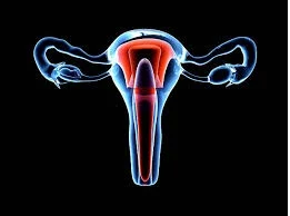
1.
INTRODUCTION:
It is a hollow muscular organ.
2.
SITUATION: -
it lies in the pelvic cavity between the urinary bladder & the rectum.
3.
SHAPE:-
pear shape or Butterfly shape.
4.
MEASURMENTS:- Length-7.5 cm long
Width- 5
cm
Thickness -2.3 cm
Weight -30- 40 grams
3 parts 1. Funds
2. Body
3. Cervix
THE FUNDUS: - this is the
dome –shaped part of the uterus above the opening of the
Uterine tubes.
THE
BODY: - this is the main part. It is narrowest inferiorly at the
internal OS where it is
Continuous with
cervix.
THE CERVIX:
- this properties through the anterior wall of the vagina opening into it at the external OS.
The walls of the uterus are composed of three
layers of issues
1.
PERIMETRIUM.
2.
MYOMETRIUM
3.
ENDOMETRIUM.
PERIMETRIUM:-
Ø This is
peritoneum which is distributed on the various surfaces of the uterus.
Ø Anteriorly it extends over the fundus & the body.
Ø Posteviorly the
petritoneum extends over the fundus.
MYOMETRIUM:-
Ø This is the
thickest layer of tissue in the uterine wall.
Ø It is a mass of
smooth muscle fiber interlaced with areolar tissue, blood vessels & nerves.
ENDOMETRIUM: -
This consists of columnar epithelium
containing a layer number of mucus secreting tubular glands. It is divided
functionally into 2 layers
1.
Upper
layer
2.
Basal
layer
UPPER LAYER:-
The functional layer upper layer &
it is thickness.
Ø It becomes rich in blood vessels in the first
half of the menstrual cycle.
Ø The basal layer
lies next to the myometrium & it is not lost during menstruation.
Ø The upper two-
third of the cervical canal is lined with this mucous membrane.
7.BLOOD SUPPLY:-
ARTERIAL SUPPLY: uterine arteries – bravely from internal iliac
arteries.
VENOUS DRAINGE: - internal iliac veins.
8.NERVE SUPPLY: - Sympathetic Nerve supply
Para Sympathetic Nerve supply.
9.LIGAMENTS OF THE UTERUS:-
1.
The
board ligaments.
2.
The
round ligaments.
3.
Uterosacral
ligaments.
4.
Transverse
cervical ligaments.
5.
Pubocervical
fascia.
10.FUNCTIONS OF THE UTERUS:-
1.
It
helps to embeds the zygote in the uterine wall.
2.
It
helps to regular monthly cycle of changes, the menstrual cycle.
3.
It
helps for the foetal growth.
11.APPLIED ANATOMY:-
1. Hysterectomy
2. Tubectomy


0 Comments