EXCRETORY SYSTEM
- The urinary system plays a vital part in maintaining homeostasis of water and electrolyte concentrations within in the body.
1. If Consists of
· 2 Kidneys - Which secret urine
· 2 ureters - which convey the urine from the kidneys to the urinary bladder.
· 1 urinary bladder – where urine collects and is temporarily stored.
· 1 urethra – through which the urine is discharged from the urinary bladder to the exterior.
- The kidneys produces the urine.
Ø Metabolic waste products
Ø Nitrogenous compounds
Ø Urea & uric acid
Ø Excessions & some drugs.,
KIDNEYS
1. INTRODUCTION:- these are the excretory organs which are present in our body.
2. SITUATION :It lie on the posterior abdominal wall, one on each side of the vertabal column, behind the peritoneum and below the diaphragm.
3. SHAPE:-“ Bean- Shaped”
4. MEASUREMENT :- Lenth - 11 cms
Wide – 6 cms
Thickness- 3 cms
Weight - 150 gms
5.ORGANS ASSOCIATED WITH THE KIDNEYS:
RIGHT KIDNEY:
SUPERIORLY: The right adrenal gland
ANTEROIRLY:The right lobe of the liver,the duodenum and the hepatic flexure of the colon.
POSTERIORLY: the diaphragm,and muscles of the posterior abdominal wall.
LEFT KIDNEY:
SUPERIORLY: The left adrenal gland
ANTEROIRLY: the spleen,stomach,pancreas,jejunum and spleenic flexure of the colon
POSTERIORLY: the diaphragm,and muscles of the posterior abdominal wall.
6.PARTS:-
1. Medulla
2. Cortex
3. Papillae of medulla,
4. Renal Pelvis.
- Fibrous Capsule:- Surrounding the Kidney
- Cortex – A reddish- brown layer of Issue
- Medulla- the innermost layer, consisting of pale conical-shaped striations of the renal pyramids.
RENAL PELVIS: it is the funnel-shaped structure which acts as a receptable for the urine formed by the kidney.
It has a no of distal branches called “Calyces”.
7.MICROSCOPIC STRUCTURE OF THE KIDNEY
1. The kidney is composed of about I million functional units, are we called it has “Nephron’s”a smaller number of collecting tubules.
2. The collecting tubules transport urine through the pyramids to the renal pelvis giving them their Stripped appearance.
3. The tubules are supported by a small amount of connective tissues,
it contains- Blood vessels, nerves & lymph vessels .
8.BLOOD SUPPLY:-
ARTERIAL SUPPLY:- RIGHT & LEFT RENAL ARTERIES
VENOUS DRIANAGE: RENAL VEIN BRANCH OF INFERIOR VENECAVA.
9. NERVE SUPPLY:-
SYMPATHETIC NERVE SUPPLY.
PARA SYMPATHETIC NERVE SUPPLY.
10.FUNCTIONS OF THE KIDNEY:
1.Formation or Urine’s
1. The kidneys from urine which passes through the ureters of the bladder for storage prior to excretion.
Composition of urine:
Water 96%
Urea 2%
 Uric acid
Creatinine
Ammonia
Sodium 2 %
Potasium
Chlorides
Phosphates
Oxalates
2. Kidneys are helps for the filtration .
3. it helps to maintain the pH level (acid-base) balance in the Body.
4. It helps for secretion
5. It helps to maintain water balance and urine output.
6. Kidneys are maintains the fluid electrolyte balance in the Body.
11.DISEASES OF THE KIDNEYS:
1. GLOMERULONEPHRITIS – Inflammation of the Glomerulus
2. ACUTE RENAL FAILURE- sudden & severu reduction in the glomerular filtration rate & kidney function.
3. RENAL CALCULI: Formation of stone in the Kidney.
URETERS
1. INTRODUCTION: The ureters are the tubes that carry urine from the kidneys to the Urinary bladder.
2. SITUATION : Behind the peritoneum & in front of the Inflammation cavity.
3. MEASUREMENTS:
Length- 25 to 30 cm long
Diameter- 3 mm
4. SHAPE:- “Funnel Shaped”
5. STRUCTURE:- 3 Layer
1. OUTER LAYER:- covering of fibrous tissue, continous with the fibrow capsule of the kidney.
2. MIDDLE LAYER:- it contains smooth muscles.
3.INNER LAYER :-it contains the mucosa ,lined with transitional epithelium.
FUNCTIONS:-
- Transmits the urine from kidney to the urinary bladder
- It produces the peristaltic movements which transmission of urine.
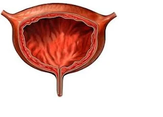
URINARY BLADDER:
1.INTRODUCTION: it is a reservoir for urine
2.SITUATION: it lies in the pelvic cavity.
3.MEASURMENTS: Its depending on the amount of urine it coulaius.
4.SHAPE: pear shaped & fills C urine it because oval shape
- organs associated with the urinary bladder
5.STRUCTURE:
IT CONSISTS 3 SURFACES
- Superior surface
- Autrior surface
- Posterior surface
the Bladder wall is Composed of 3 layers
OUTER LAYER:-
- layer of loose connective tissues,
- It contains Blood & Lyuphenodes & nerves
- It covered on the upper surface by the peritneum
MIDDLE LAYER:
It contains smooth accucle fibres & elastic tissue,
The mucosal –lined with Transitional epithelium.
Capacity of the urinary bladder 300-400ml urinate initiated.
Total capacity is rarely more than 600ml.
FUNCTIONS:
Storage (or) reserviour for the urine.
URETHRA
The urethra is a canal extending from the neck of the bladder to the exterior.
Its length differs in the male compare to the female.
THE FEMALE URETHRA is approximately 4cm long.
It runs downwards and forwards behind the Symphysis pubis and opens at the external urethral orifice just in front of vagina.
It is under voluntary control.
THE MALE URETHRA:
It provides a common pathway for the flow of urine and Semen.
It is about 19 to 20 cm long.
It is also under voluntary control.
It consists of 3 layers.
1. The muscular layer.
2. Sub mucosa layer.
3. Mucosa layer.


0 Comments