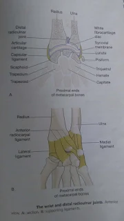
WRIST JOINT
v
This is a
condyloid joint between the distal end of the radius and the proximal ends of
the scaphoid, lunate and triquetral.
v
A disc of
white fibrocartilage separates the ulna from the joint cavity and articulats
with the carpal bones.
v
It also
separates the inferior radioulnar joint from the wrist joint.
v
Extracapsular
structures consist of medial and lateral ligament and anterior and posterior
radiocarpal ligaments.
MUSCLES AND MOVEMENTS
1. The wrist can be flexed, extended, abducted
and adducted.
2. The muscles that perform these movement are
the main muscles that move the wrist.
JOINT OF THE HANDS AND
FINGERS
1. There are synovial joints between the
carpal bones, between the carpal and metacarpal bones, between the metacarpal
bones and proximal phalanges and between the phalanges.
2. Movement at the hand and finger joints are
controlled by muscles in the forearm and smaller muscles within the hand.
3. There are no muscles in the fingers; finger
movements are produced by tendons extending from muscles in the forearm and the
hand.
4. The joint at the base of the thumb is a
saddle joint, unlike the corresponding joints of the other fingers, with are
condyloid.
5. This means that the thumb is more mobile
than the fingers and the thumb can be flexed, extended, circumducted, abducted
and adducted.
6. In addition, the thumb can be moved across
the palm to touch the tips of each of the fingers on the same hand
(opposition), an ability that confers great manual dexterity and allows, for
example, the holding of a pen and the fine manipulation of objects held in the
hand.
7. The joints between the wrist and finger
bones allow movement of the fingers.
8. The fingers may be flexed, extended,
adducted, abducted and circumducted, with the first finger having the greatest
flexibility of movement.
9. The finger joints are hinge joints, and
allow only flexion and extension.
10. The flexor retinaculum is a strong fibrous
band that stretches across the front of the carpal bones, enclosing their
concavity and forming the carpal tunnel.
11. The tendons of flexor muscles of the wrist
joint and the fingers and the medial nerve pass through the carpal tunnel, the
retinaculum holding them close to the bones.
12. Synovial membrane forms sleeves around
these tendons in the carpal tunnel and extends some way into the palm of the
hand.
13. Synovial sheaths also enclose the tendons
on the flexor surfaces of the fingers.
14. Their synovial fluid prevents friction that
might damage the tendons as they move over the bones.
15. The extensor retinaculum is a strong
fibrous band that extends across the back of the wrist.
16. Tendons of muscles that extend the wrist
and finger joints are encased in synovial membrane under the retinaculum.
17. The synovial sheaths are less extensive
than on the flexor aspect.
18. The synovial fluid secreted prevents
friction.


0 Comments