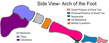
TARSAL (ANKLE) BONES:

The seven tarsal bones forming the posterior part of the
foot (ankle) are the talus, calcaneus, navicular, cuboid and three cuneiform
bones.

The talus articulates
with the tibia and fibula at the ankle joint.
The calcaneus forms the heel of the foot.
The other bones
articulate with each other and with the metatarsal bones.
METATARSALS (BONES OF THE FOOT):

There are five bones, numbered form inside out, which form
the greater part of the dorsum of the foot.

At their proximal ends they articulate with the tarsal bones
and at their distal ends, with the phalanges.

The enlarged distal head of the 1st metatarsal
bone forms the ‘ball’ of the foot.
PHALANGES (TOE BONES):

There are 14 phalanges arranged in a similar manner to those
in the fingers, i.e. two in the great toe (the halux) and three in each of the
other toes.

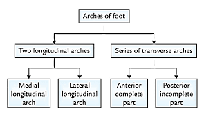
ARCHES OF THE FOOT.

The arrangement of bones in the foot, supported b y
associated ligaments and action of associated muscles, gives the sole of the
foot an arched or curved shape.
The curve running
from heel to toe is called longitudinal arch, and the curve running across the
foot is called the transverse arch.
In the normal longitudinal arch, only the calcaneus and the
distal ends of the metatarsals should touch the ground, the bones in between
lifted clear.
This gives a conventional footprint shape.
If, however, the
concavity of the sole is lost because of sagging ligaments or tendons, the arch
sinks and much more of the sole of the foot is in contact with the ground; this
is called flat foot.
Because the arches of
the foot are important in distributing the weight of the body evenly whilst
upright, whether stationary or moving, the flat foot loses the springiness of
normal foot structure and leads to sore feet when standing, walking or running
for long periods.
As there are movable
joints between all the bones of the foot, very strong muscles and ligaments are
necessary to maintain the strength, resilience and stability of the foot during
walking, running and jumping.
POSTERIOR TIBIALIS MUSCLE.
This is the most
important muscle support of the longitudinal arch.
It lies on the posterior
aspect of the lower leg, originates from the middle third of the tibia and
fibula and its tendon passes behind the medial malleolus to be inserted into
the navicular, cuneiform, cuboid and metatarsal bones. It acts as a sling or
‘suspension apparatus’ for the arch.
SHORT MUSCLES OF THE FOOT.
This group of muscles
is mainly concerned with the maintenance of the longitudinal and transverse
arches. They make up the fleshy part of the sole of the foot.
PLANTAR CALCANEONAVICULAR LIGAMENT
(‘SPRING’ LIGAMENT).
This is very strong
thick ligament stretching from the calcaneus to the navicular bone. It plays an
important part in supporting the medial longitudinal arch.
Plantar ligaments and interosseous membranes. These
structures support the lateral and transverse arches.


 BONES:)
0 Comments