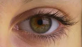
EYE
1.INTRODUCTION:
The eye is the organ of the sense of sight or vision .
2.SITUATION:
It
is situated in the orbital cavity.
3.MEASURMENTS:
v The space between eye and the orbital cavity is occupied by adipose
tissue.
v The bony walls of the orbit and the fat help to protect the from injury.
4.ACCESSORY ORGANS OF THE EYE:
The eye is a delicate organ which is
protected by several structures.
Eye brows
Eye lids and eye lashes
Lacrimial apparatus.
5.STRUCTURE:
There are 3 layers of tissue in the walls of
the eye
1.The outer fibrous layer : sclera
& cornea
2.the middle vascular layer (or)
uveal tract :choroid ciliary body & iris.
3. the inner nervous tissue layer :
retina.
4.structures inside the eyeball are
the lens ,aqueous fluid (humour) & vitreous
body (humour).
SCLERA & CORNEA:
THE
SCLERA
- It is a white part of the eye.
- It forms the outermost layer of tissue of the posterior and lateral aspects of the eye ball,is continous anteriorly with the transparent cornea.
- It consists of a firm fibrous membrane that maintains the shape of the eye and gives attachment to the extraocular (or) extrinsic muscles of the eye.
- Anteriorly the sclera continues as a clear transparent epithelial membrane, the ‘cornea’.
- Light rays pass through the cornea to reach the retina.
- The cornea is convex anteriorly and is involved in refracting or bending light rays to focus them on the retina.
CHOROID:
- The choroid lines the posteriorly five-sixth of the inner surface of the sclera.
- It is very rich in blood vessels and is deep chocolate brown in colour.
- Light enters the eye through the pupil , stimulates the nerve endings in the retina and is then absorbed by the choroid.
CILIARY
BODY:
- The ciliary body is the anterior contituation of the choroid consisting of ciliary muscle and secretory epithelial cells.
- It gives attachment to the sensory ligament which at its other end is attached to the capsule enclosing the lens.
- Contraction and relaxation of the ciliary muscle changes the thickness of the lens which bends or refracts light rays entering the to focus them on the retina.
- The epithelial cells secrete “Aqueos fluid “ into the anterior segment of the eye,I,e the space between the lens and the cornea (anterior & posterior compartment.
- The ciliary body is supplied by parasymphathetic branches of the oculo motor nerve(3rd cranial nerve).
- Stimulation causes contraction of the smooth muscle and accommodation of the eye.
IRIS:
- The iris is the visible coloured part of the eye & extends anteriorly from the ciliary body,lying behind the cornea infront of the lens.
- The colour of the iris is genetically determined and depends on the number of pigment cells present.
- Albinos have no pigment cells and people with blue eyes have fewer then those with brown eyes.
- It divides the anterior segment of the eye into anterior and posterior chambers which contains “Aqueous fluid” secreted by the ciliary body.
- It is a circular body composed of pigment cells and two layers of smooth muscle fibres, one circular and other radiating.
- In the centre there is an aperture called the PUPIL.
- The iris is supplied by parasymphathetic and parasymphethetic nerves.
- Parasympathetic nerve stimulation constricts the pupil and sympathetic stimulation dilates the pupil.
LENS
- The lens is a highly elastic circular biconvex body,lying immediately behind the pupil.
- It consists of fibres enclosed within a capsule and it is suspended from the ciliary body by the suspensory ligament .
- The lens bends (refracts) light rays relected by objects in front of the eye.
- Lens is the structure in the eye that can vary its refractory power,achived by changing its thickness.
- When the ciliary muscle contracts, it moves forward,releasing its pull on the lens, increasing its thickness.
- The nearer is the object being viewed the thicker the lens becomes to allow focusing.
RETINA:
- The retina is the innermost layer of the wall of the eye.
- It is an extremely delicate structure and is especially adapted for stimulation by light rays.
- It is composed of several layers of nerve cell bodies and their axons, lying as a pigmented layer of epithelial cells which attach it to the choroid.
- The layer highly sensitive to light is the layer of sensory receptor cells :rods & cones.
- The retina lines about three-quarters of the eyeball.
- The eyeball is thickest at the back and things out anteriorly to end just behind the ciliary body.
- Near the center of the posterior part is the macculalutea, (or) yellow spot.
- In the centre of the area there is a little depression called “fovea centralis”. Consisting of only ‘cone-shaped cells.
- The rods and cones contain photosensitive pigments that convert light rays into nerve impulses.
- About 0.5 cm to the nasal side of the macula lutea all the nerve fibres of the retina converge to form the optic nerve.
- The small area of the retina where the optic nerve leaves the eye is the optic disc (or) blind spot.
- It has no light sensitive cells.
6.BLOOD SUPPLY:
ARTERIAL
BLOOD SUPPLY:
- Cliary artery and thr central retinal artery these are the branches of ophthalmic artery and branches of the carotid artery.
VENOUS
DRAINAGE:
- Central retinal vein which eventually empty into a deep venous sinus.


0 Comments