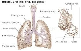
1. INTRODUCTION:- “ A Bronchus also brown as a main (or) primary bronchus, is an airway in the respiratory tract that conducts air into the lungs.
2. SITUATION: - The Level of the 5th thoracic Vertebra
3. PARTS:-
The Right Bronchus.
The Left Bronchus.
1. This is wider, shorter & more vertical than the lt bronchus & is therefore more likely to become obstructed by an inhaled foreign body.,
2. Length:- Approximately-2.5 cm long.
3. After entering the Rt lung at the hilum it divides into three branches Out to each lobe.
LT BRONCHUS:-
1. This is about 5cm long & is narrow than the right.
2. After entering the lung at the hilum it divides into two branches, one to each lobe ,
4. STRUCTURE:-.
· The bronchi are composed of the same tissues as the Trachea, and are lined with ciliated columnar epithelium.
· The bronchi subdivided into branchioles, terminal bronchioles, respiratory bronchioles, alveolar ducts & aveoli.
· The distal end of the bronchi the cartilages become irregular in shape and are absent at bronchiolar level.
· In the absence of cartiliage the smooth muscle in the walls of the bronchioles became thicker & is responsive to autonomic nerve stimulation & irritation.
· Ciliated coloumnar mucus memberane changes gradually to non-ciliated cuboidal-shaped calls In the distal bronchioles.
· The wider passage are called conducting airways.
· Conducting airways function is to bring air into the lungs & their walks are too thick to permit gas exchange.
5. BLOOD SUPPLY:-
Arterial Supply :- the Rt & Lt Bronchial arteries.
Venous Returns:- Bronchial veins.
6. NEVER SUPPLY:- Parasympathetic:- Vagus nerve Stimulates contraction of smooth muscle causes “ Branchoconstriction. Sympathetic nerve supply: Stimulation causes Bronchodilation.
7. LYMPH SUPPLY:- Lymph is drained from the walls of the air passages in a network of lymph vessels


0 Comments