HEART
1.
INTRODUCTION:
The heart
is the roughly hollow muscular organ.
2.
SITUATION:
Ø The heart lies in the thoracic cavity in the
mediastinum between the lungs.
Ø It lies obliquely a little more to the left than the
right and presents a base above & an apex below.
3.
MEASURMENTS:
Ø SHAPE
- CONE SHAPE
Ø LENGTH
- 10 cm long
Ø SIZE - 225 gm in women.
More than 225 gm
in Men.
4.
ORGANS ASSOCIATED WITH THE HEART:
Ø Inferiorly – the apex rests on the central trends f
the diaphragm.
Ø Superiorly- Blood venal that is the aorla, SVC,
Pulmonary artery & Pulmonary veins.
Ø Portiorly - The Oesophagus, trachea, left & right broumnchues, m Desceneling aorla, IVC & thoracic vertebrae.
Ø Laterally -
The lungs the left lung overlaps the left side of the heart.
Ø Anterior -
The sterum, Ribs & Intercostals muscles.
5.
STRUCTURE:
The heart is composed of 3 layers
Ø Pericardium
Ø Myocardium
Ø Endocardium
PERICARDIUM:
It
is the outer conversing layer of the heart, made up of 2 sacs.
Ø Other sac
- commits fibrous tissue
Ø Inner sac -
Double layer of Serous membrane.
The outer fibrous sac is
continuous. With the tunica adventitia of the great blood vessels above and is
adherent to the diaphragm below.
Ø It’s elastic ,
fibrous nature.
Ø The outer layer of the serous membrane, the parietal
pericardium, lines the fibrous sac.
Ø The Inner layer, the visceral pericardium or
epicardium, which is continuous with the parietal pericardium, is adherents to
the heart muscle.
Ø Serous membrane consists of flattened epithelial
cells.
Ø It secretes serous third into the space between the
visceral &parietal layers which allows smooth movement between them when
the heart beats.
Ø The between the parietal & visceral pericardium
is only a potential space.
MYOCARDIUM:
The Myocardium is composed of
specialized cardiac muscle found only in the heart.
Ø It is not
under voluntary control but like skeletal muscle, cross stripes are seen on
microscopic examinations.
Ø Each fiber has nuclei and one or more branches.
Ø Microscopically these Joints or Intercalated discs
can be seen as thicker,darker lines than the ordinary cross stripes.
Ø When an impulse is initiated. It spreads from cell
to cell via the branches and intercalated discs over the whole sheet of muscle,
causing contraction.
Ø The sheet arrangement of the myocardium enables the
atria & ventricles to contract in a coordinated & efficient manner.
Ø The myocardium is thickest at the Apex & thins
out towards the base.
Ø This reflects the amount of work each chamber
contributes to the pumping of blood.
Ø It is thickest in the left ventricle.
Ø The Atria & the ventricles are separated by a
ring of fibrous tissue that doesn’t conduct electrical impulses.
ENDOCARDIUM: -
It forms the
lining of the myocardium and the heart values.
Ø It is a thin, smooth, glistening membrane which
permits smooth flow of blood inside the heart.
Ø It consists of flattened epithelial cells,
continuous with the endothelium that lines the blood verses.
INTERIOR
OF THE HEART:
The heart is
divided into a right & left side by the septum, a partition consisting of
myocardium covered by endocardium.
Ø After birth blood can’t cross the septum from one
side to the other.
Ø Each side is divided by an Atrioventricular value
into an upper chamber, the Atrium and a lower chamber the ventricle.
Ø The Atrioventricular values are formed by double
folds of endocardium strengthened by a little fibrous tissue.
Ø The Retro ventricular value [Tricuspid value] has 3
flaps or cusps & the it atrioventricular value or Bicuspid has two cusps.
Ø The values between the atria & ventricles open
& close passively according to change in pressure in the chambers.
Ø They open when the pressure in the Atria is greater
than that in the ventricles.
Ø During ventricular systole (contraction) the
pressure in the ventricles reissues above that in the Artia & the values
snap that preventing backward flow of blood.
Ø The values are prevented from opening upwards into
the atria by tendinous cords called ‘ Chordate lendineae’ which expends from the inferior surface of the cusps to little projections of myocardium covered with endothelium called
‘papillary muscles’.
6.
BLOOD
SUPPLY
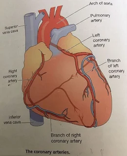
ARTERIAL SUPPLY
The right & left
coronary oratories which branches from the aorta immediately distal to the
aortic value.
VENOUS DRAINAGE:
The
venous blood is collected into several small veins that join to from the
coronary sinus which opens into the right Atrium.
7.
NERVE
SUPPLY:
PARASYMPHATHETIC N S:
The
Vegas nerves supply mainly the SA & AV nodes and aerial muscles
Ø It reduces the Impulses & Decreases the heart
rate
SYMPATHETIC N S:
It supplies the SA & AV nodes & the myocardium of Atria & Ventricles.
Ø Sympatyhic stimulation increase the rate & force
of the heart beat.
8.
FACTORS
AFFECTING HEART RATE:
1.
AUTOMATIC
NERVOUS SYSTEM:
The
rate at which the heart beat is a balance of sympathetic & parasympathetic
activity.
2.
CIRCULATING
CHEMICALS:
The hormones Adrenaline & nor adrenaline
secreted by the adrenal medulla, it increase the heartrate.
3.
POSITION:
When
the person is upright the heart rate is usually faster than when lying down.
4.
EXERCISE:
Active
muscles med more blood than resting muscles &this is achieved by an
increased heart rate & selective vasodilatation.
5.
EMOTIONAL
STRESS:
During
excitement fear or anxiety the heart rate is increased.
6.
GENDER:
The
heart rate is faster in women than men.
7.
AGE:
In a babies & small children the heart
rate is more rapid than in older children & adults.
8.
TEMPERATURE:
The heart rate increases & decreases with
body temperature.
9.
BARORECEPTOR
REFLEX:
These
are nerve endings sensitive to pressure changes ( stretch) within the vessel
situated in the Arch of the aorta & in the carotid sinuses.


0 Comments